

This new section reviews the theory and provides real-world examples of clinical application of PCCT.
All you need to know about PCCT

CHRISTIAN LOEWE
Division of Cardiovascular and Interventional Radiology
Dept. of Bioimaging and Image-Guided Therapy
Medical University Vienna
Photon-counting computed tomography (PCCT) is a new CT technology that utilises a direct conversion X-ray detector, in which the energy of the incident X-ray photon is directly recorded as electronical signal.
There are several advantages to PCCT over the traditional CT scanners incorporating energy-integrating detectors (EIDs). These include improvements in spatial resolution (via smaller detector pixel design) and iodine signal (via count weighting), still permitting multi-energy imaging, electronic noise elimination, and artefact reduction (via energy thresholds).

PCCT Clinical Cases

Thom van der Laan, Judith van der Bie, Ricardo Budde, Zeen Aref
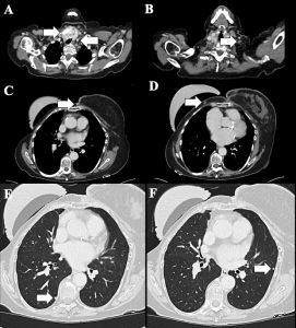
PCCT in the follow up of treatment for angiosarcoma
A 71-year-old female patient with a history of right breast carcinoma (1984) treated with modified radical mastectomy (GRM), chemotherapy, and radiotherapy.
Judith van der Bie, Thom van der Laan, Ricardo Budde, Zeen Aref
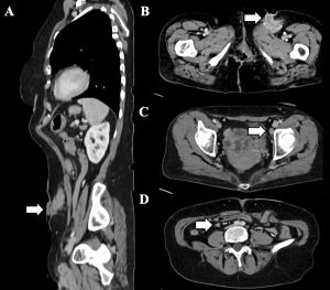
PCCT in the diagnosis of recurrent vulvar tumour
A 74-year-old woman with vulvar carcinoma requiring posterior exenteration and reconstruction due to disease extent was suspected of having a recurrent tumour in the left groin.

Judith van der Bie, Simran Sharma, Thom van der Laan, Alexander Hirsch, Ricardo Budde
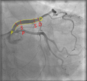
PCCT in the follow-up of percutaneous coronary intervention – stent patency
A 60-year-old woman with recurrent angina who underwent percutaneous coronary intervention with stent placement in the left anterior descending artery in 2022.
Judith van der Bie, Simran Sharma, Thom van der Laan, Alexander Hirsch, Ricardo Budde
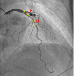
PCCT in the follow-up of percutaneous coronary intervention – restenosis
A 59-year-old man with recurrent angina underwent percutaneous coronary intervention (PCI) with stent placement in the left anterior descending artery (LAD) in 2004.
Lucian Beer
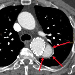
PCCT in the follow-up of aortal stenting (3)
A 66 year-old male patient was referred for routine follow up after type B aortic dissection treated with a branched thoracoabdominal stent-graft 8 months earlier. The patient was asymptomatic at time of imaging.
Lucian Beer
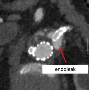
PCCT in the follow-up of aortal stenting (2)
An 84-year-old male patient was referred for routine follow-up after four-times branched endovascular repair (EVAR) of an infrarenal aortic aneurysm. The patient was asymptomatic at the time of imaging.
Lucian Beer
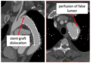
PCCT in the follow-up of aortal stenting (1)
A 44-year-old male patient was referred for routine follow-up after type B aortic dissection treated with a thoracic stent-graft 10 months earlier. The patient was asymptomatic at the time of imaging.
Lucian Beer
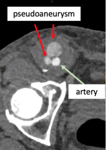
PCCT in the diagnosis of a complex pseudoaneurysm
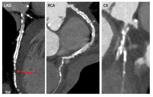
A 60-year-old male patient was referred for further workup due to a pseudoaneurysm in the right groin after bypass surgery of the right common femoral artery two weeks earlier.
Lucian Beer
PCCT in the diagnosis of a suspected mass between atria
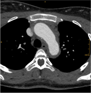
A 78-year-old male patient was referred for further workup owing toa suspected mass between the right and left atrium seen on cardiac echography. He had known stable ischemic cardiomyopathy.
Lucian Beer
PCCT in the diagnosis of the Takayasu arteritis
A 22-year-old woman (59 kg) with juvenile idiopathic arthritis (JIA) and uveitis was referred to rule out large vessel vasculitis. At the time of imaging she was asymptomatic in terms of JIA.
Lucian Beer
PCCT in the diagnosis of Pancreatic Neuroendocrine Tumour
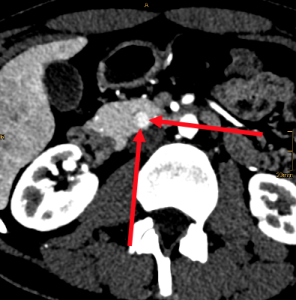
A 25-year-old asymptomatic female patient was referred to rule out an endocrine neoplasm of the pancreas given her positive family history of neuroendocrine neoplasm and MEN1 mutation.
Lucian Beer
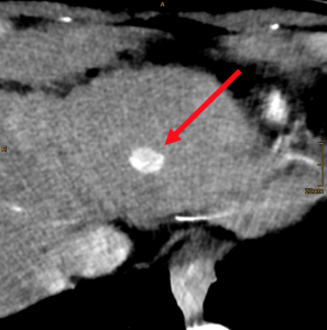
PCCT in the diagnosis of Clear Cell Renal Cell Carcinoma
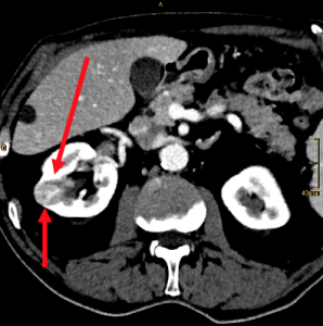
A 72-year-old male patient was referred for further workup due to a suspected right kidney tumour. He presented no symptoms, and there was no family history of cancer.
Lucian Beer
PCCT in the diagnosis of Hepatocellular Carcinoma
A 60-year-old male patient with advanced chronic liver disease due to chronic hepatitis B infection was referred due to suspicion of hepatocellular carcinoma (HCC) in the left liver lobe.
Lucian Beer
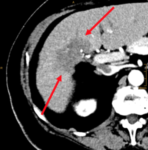
PCCT in the diagnosis of Intrahepatic Cholangiocellular Carcinoma
A 70-year-old female patient was referred for suspected cholangiocellular carcinoma workup. She presented with diffuse abdominal pain but no further clinical signs.