Dear readers,
Abdominal wall hernias comprise a heterogeneous group of entities that differ in location, size, content, origin, and underlying etiology. Contrast-enhanced CT is widely recognized as the most reliable imaging modality for their evaluation, as it provides accurate anatomical depiction and supports the differential diagnosis.
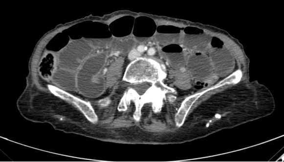
In the videopill featured in this Newsletter, Dr. Perez-Girbes (Hospital Universitari i Politècnic La Fe, Valencia, Spain) illustrates the role of CT in distinguishing primary (congenital or acquired) from incisional hernias, in accordance with the European Hernia Society classification. The discussion also touches on inguinal hernias, on account of their high prevalence. The videopill is enriched by several illustrative CT cases highlighting the key imaging features required for accurate detection and characterization.
Two selected scientific articles further emphasize the value of CT imaging in this context: the first explores morphometric changes to the abdominal wall following reconstruction with the transversus abdominis release technique in patients with incisional ventral hernia, while the second highlights the role of CT in redefining the anatomical basis of the umbilical canal, often simplistically referred to as “umbilical hernia” in radiology reports.
Finally, a clinical case underscores the contribution of CT imaging in the differential diagnosis of the various types of inguinal hernias.
As always, we welcome suggestions for topics to be addressed in future issues of this Newsletter: please feel free to contact us at editorial@mdct.net.
From the MDCT.net team